

Biomechanics Laboratory
 Research groups, Dr. Yucesoy had contributed to, showed in the last decade that substantial amounts of muscle force can be transmitted via non-tendinous pathways comprised of direct collageneous connections between neighboring muscles and neurovascular tracts in continuity with compartment delimiting connective tissues. The latter provides connections between not only synergistic muscles but also between antagonistic muscles as well as between muscle and non-muscular structures. Experimentally and using finite element modeling, such epimuscular myofascial force transmission has been shown to affect muscular mechanics in a major way: (1) proximo-distal muscle force differences, (2) mechanical condition dependent muscle length-force characteristics and (3) major sarcomere length heterogeneity are the characteristic effects obtained in situ. The goal of this work is to show, using MRI coupled with DTI that epimuscular myofascial force transmission has major effects also in human muscles and in vivo: measurement of human muscle force in vivo is very difficult if not impossible however, using such imaging, calculation of local muscle tissue deformations in the direction of muscle fibers occurring after joint motion is possible. This will allow us to achieve a representation of sarcomere length distributions which our research shows that is a key parameter for improving our understanding of many muscle diseases and their treatment. The current project involves:
 .
. 
Biomechanics Laboratory
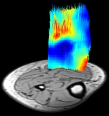
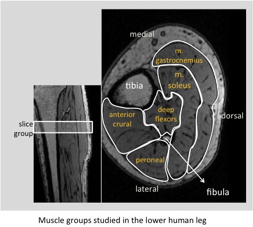
Due to myofascial loads, strain distribution within muscle cannot be homogeneous. Using MRI based deformation analyses and DTI based tractography combined, we aimed at assessing muscle fascicle direction local tissue deformations within the human medial gastrocnemius muscle as caused by knee angle changes. Tractography and Demons nonrigid registration algorithm were utilized to calculate local deformations along muscle fascicles. Despite global lengthening conditions, fascicle tracts with negative and positive mean strains exist. Much shearing across fascicle tracts have been observed. Non-uniformity of fascicle strain indicates epimuscular myofascial force transmission. MRI and DTI analyses combined provide a powerful tool for quantifying deformation along human muscle fibers in vivo.
